Integrated rock analysis for unparalleled subsurface insight
talk to an expertIntegrated rock analysis provides the link between downhole measurements and physical properties of the reservoir, including those that control fluid flow. This information can be used to improve well placement, wellbore management, completion design, and recovery rates.
Integrated rock analysis solutions, has led the market with commercial-scale workflows for integrated rock analysis in core, plugs, sidewall cores, cuttings, rock chips, and thin sections. We provide these services worldwide with laboratories in South America and the Middle East.
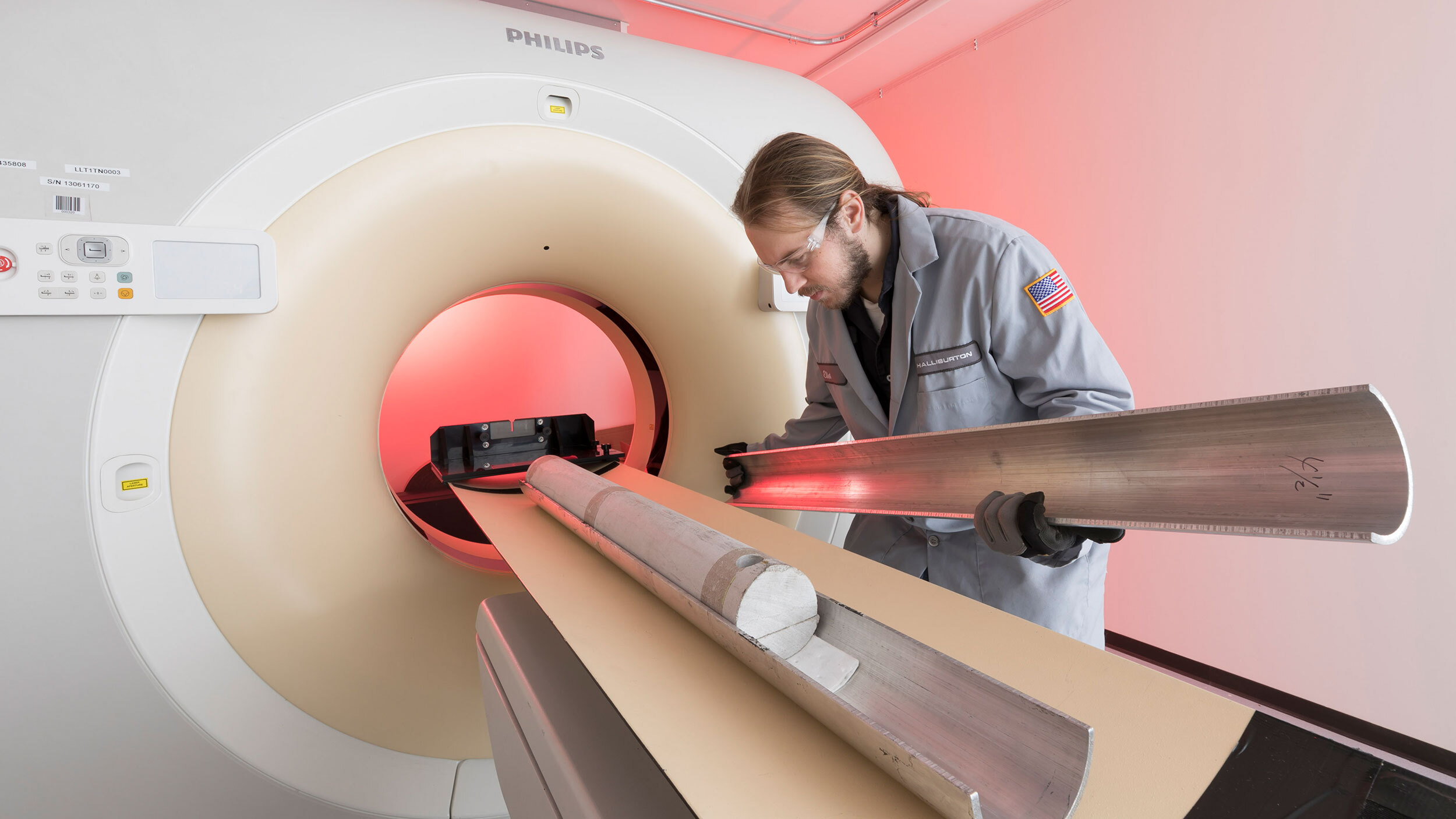
We combine state-of-the-art digital imaging, computational fluid dynamics, and physical laboratory measurements to deliver and upscale routine and special core analysis properties in an accelerated timeframe. Our digital imaging technology includes methods for dual energy CT, micro- and nano-CT, and focused ion beam scanning electron microscopy (FIB-SEM). This technology produces images and data that are used to define static and dynamic rock properties, which can be upscaled from pore to core scale and then integrated with well logs.
We are pioneering workflows for sample preparation and imaging of whole/slabbed cores, plugs, sidewall cores, cuttings, rock chips, and thin section micrographs, down to the pore scale. integrated rock analysis solutions, will accommodate special imaging protocols and sample-preparation requests from our customers.
South America and Middle East
Sampling reduction with CorePoint®
Multi-resolution imaging from 300 μm to 1 nm
More than 35 patents in digital rock analysis